- About ASOPRS
- Membership
- Symposia
- Fellowships
- OPS Community
Blocked Tear Ducts – Adults Dacryocystorhinostomy (DCR)
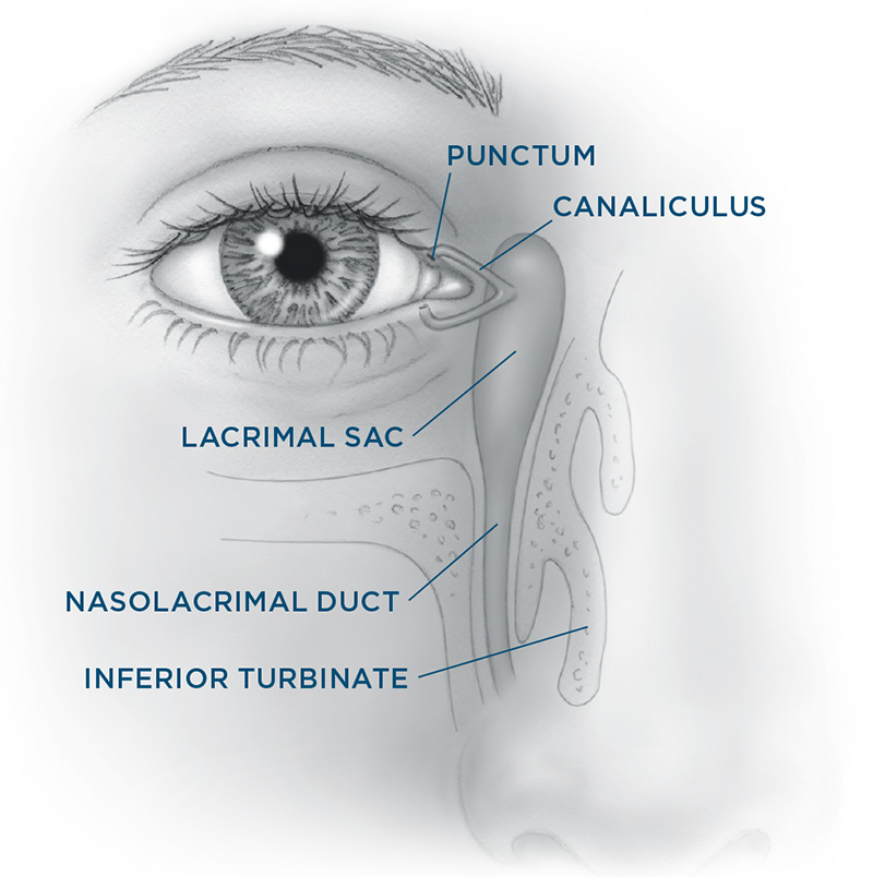
When you blink, your eyelids push tears evenly across the eyes to keep them moist and healthy. Blinking also pumps your old tears into the puncta and lacrimal sac where they travel through the nasolacrimal duct and drain into your nose. The most common symptoms include mucous buildup at the inside corner of the eye and/or along the lashes, excessive watering and distorted vision. Depending on where along the passageway from the punctum to the nose the blockage occurs, you may also have redness, tenderness and swelling between the inner corner of the eye and the side of the nose. A skillful history and examination by your doctor can usually identify if a narrowing or blockage exists and if so, where along the passageway it is. Treatments
To perform the procedure, your surgeon will create a new drainage opening from the blocked sac directly into your nose to bypass the obstruction in your nasolacrimal duct. (Unlike children in whom a blocked nasolacrimal duct can often be opened with a sharp probe, adults are not able to be treated in this same way.) A small incision is made either in the skin or inside the nose. A fine, soft silicone stent may temporarily be left in the new tear drain for a few weeks to keep the duct open while healing occurs. A DCR is an outpatient procedure that may be done under “twilight” sedation or general anesthesia. You may have a bit of nose bleeding for a couple days after the procedure. Most people recover in less than a week. A less common site of complete blockage is at the level of the canaliculus. In this situation, a prosthetic Pyrex glass prosthesis (often referred to as a Jones or Gladstone-Putterman tube) may be placed to connect the surface of the eye directly to the nasal cavity in a procedure called a conjunctivodacryocystorhinostomy (CDCR). Unlike a DCR that requires virtually no maintenance after surgery, living with a glass tube can create challenges for many patients. While some patients may choose to simply live with their tearing in this situation, your surgeon can provide more details to help you make your decision. Most patients experience improvement in their tearing and discharge once the appropriate procedure has been done to address the blockage in their tear drainage system. Risks and Complications Your surgeon cannot control all the variables that may impact your final result. The goal is always to improve a patient’s condition but no guarantees or promises can be made for a successful outcome in any surgical procedure. There is always a chance you will not be satisfied with your results and/or that you will need additional treatment. As with any medical decision, there may be other inherent risks or alternatives that should be discussed with your surgeon. Blocked Tear Ducts - Adults Dacryocystorhinostomy (DCR) Photos • Find an ASOPRS Surgeon Near You |

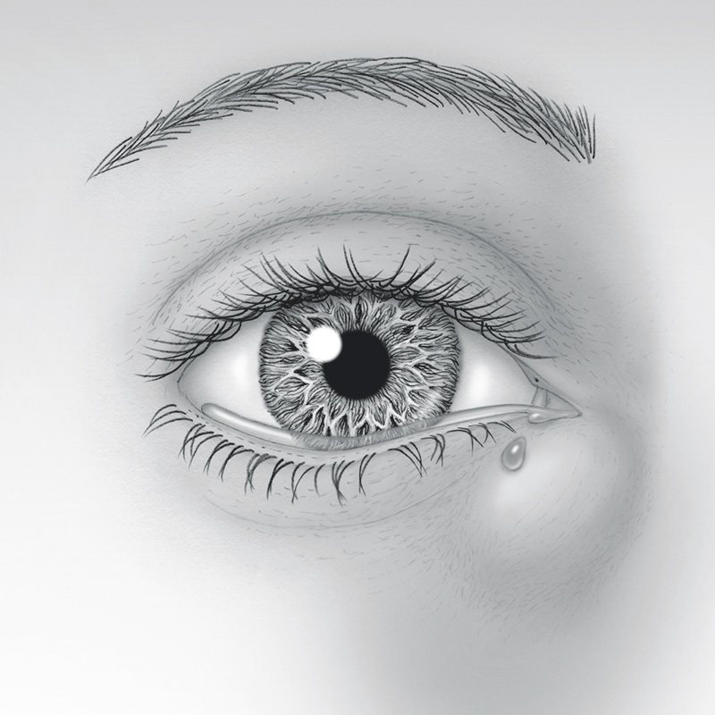
 The tear drain starts with two small openings called puncta; one punctum is in your inner upper eyelid and the other is in the inner lower eyelid. You can see them with the naked eye if you look up close. Each of these openings leads into a small tube called the canaliculus which in turn empties into the lacrimal sac between the inside corner of your eye and nose. The lacrimal sac narrows into a tunnel called the nasolacrimal duct that passes through the bony structures surrounding your nose and then empties tears into your nasal cavity.
 Narrowing of the punctum or canaliculus may respond to minor office procedures to reopen these passageways — snip punctoplasty or stenting procedures. A common location of blockage is the nasolacrimal duct. This causes tears to be trapped in the lacrimal sac and sometimes become stagnant and infected. If the nasolacrimal duct is narrowed (also known as “stenosis”) but still partially open, your surgeon may recommend placing temporary stents through your nasal passageways. If this is not effective or if the nasolacrimal duct becomes completely blocked, a dacrocystorhinostomy (DCR) is the gold standard surgery to correct this problem.